Three-dimensional time of flight magnetic resonance angiography of the heart and associated vessels in a cat
Journal of Veterinary Cardiology,Volume 18, Issue 4, December 2016, Pages 413–417
Abstract
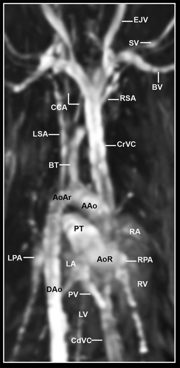
The aim of this study was to describe the normal magnetic resonance angiography (MRA) of the heart and associated vessels in a mature female cat using a 1.5-Tesla magnet. Non-contrast enhanced MRA was performed using a three-dimensional time of flight (TOF) sequence in parasagittal and dorsal aspects. Relevant cardiac and vascular structures were labelled on three-dimensional Time of flight images. Time of flight imaging showed details of the heart cavities and vessels lumen due to the high signal intensity of fast-flowing blood compared with bones, muscles, and lungs, which appeared with low signal intensity. Three-dimensional TOF sequences provided adequate anatomical details of the heart and good differentiation of the vascular structures that could be used for interpretation of cardiac images and to assist in future MRA studies.
Enlace al Artículo
