Computed tomography of the brain and associated structures of the one-humped camel (Camelus dromedarius): An anatomic study
Journal of Applied Animal Research. Volume 43, Issue 2, 3 April 2015, Pages 218-223
Blanco D, Vázquez JM, Rivero, M.A., Corbera, J.A., Arencibia, A.
Abstract
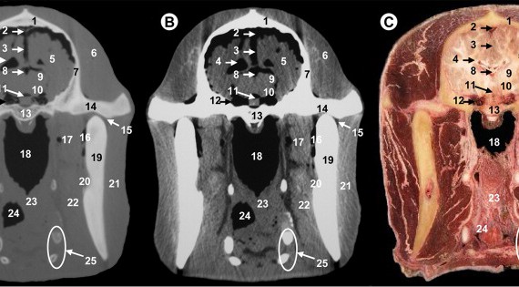
The purpose of the study was to provide a description of normal cross-sectional anatomy of the camel brain and associated structures using computed tomography (CT) and macroscopic cross sections. Transverse images of two isolated camel cadaver heads were obtained by an axial CT equipment. CT scans were processed with a detailed algorithm using bone and soft-tissue windows settings, and compared with the corresponding frozen cross sections of the heads, to assist in the accurate identification of brain and associated structures. CT images provided good differentiation between the bones and the soft tissues of the head. These CT images are intended to be a useful anatomic reference in the interpretation for clinical CT imaging studies of the brain and associated structures in dromedary camels.

